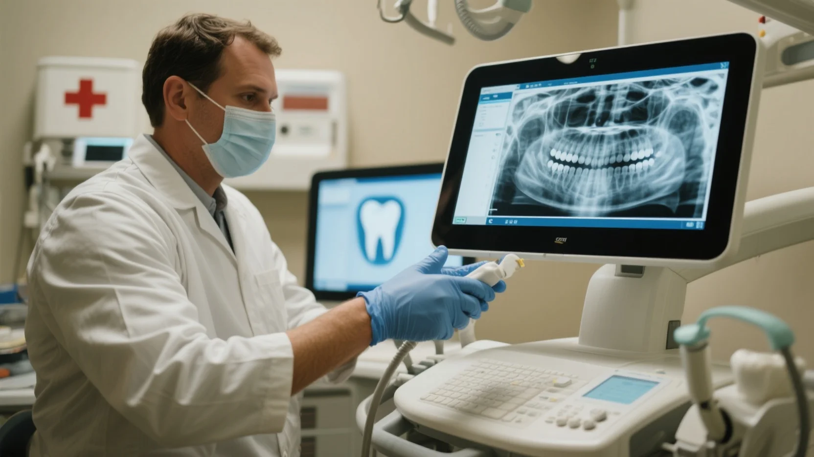
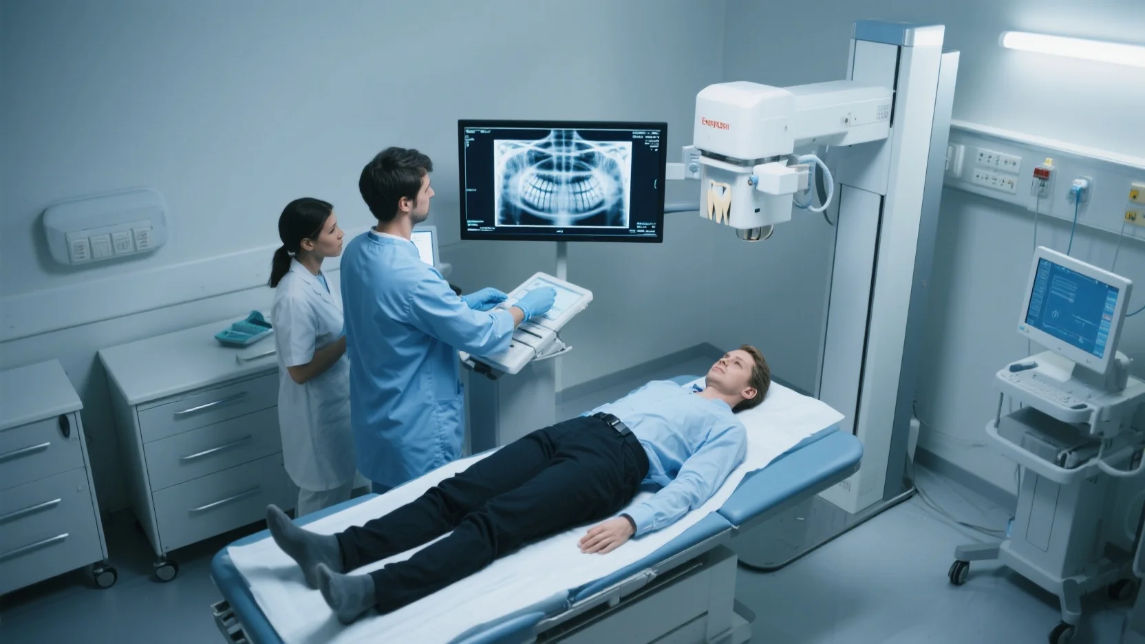
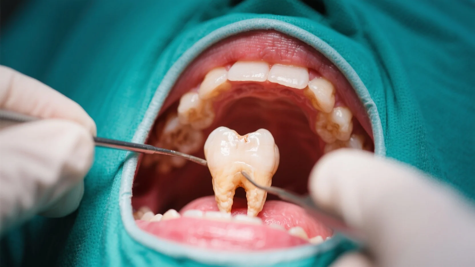
In the United States, where someone visits a hospital emergency department for dental conditions every 15 seconds, emergency dental X – ray services are a critical necessity. According to a SEMrush 2023 Study and the American Dental Association, these X – rays can cost from $50 to over $1000, depending on the type. Premium emergency X – rays offer high – resolution images for accurate diagnosis compared to counterfeit – like, inaccurate results from subpar providers. With our Best Price Guarantee and Free Installation Included for some on – site dental imaging setups, don’t miss out on getting the right urgent radiographic evaluation now!
Cost
Did you know that in the United States, on average, someone visits a hospital emergency department for dental conditions every 15 seconds? Understanding the cost associated with emergency dental X – rays is crucial for patients, providers, and insurers alike.
Range of costs
The cost of emergency dental X – rays can vary significantly. According to industry data, basic dental X – rays in an emergency setting can range from $50 to $200. However, more complex panoramic or cone – beam CT scans can cost anywhere from $250 to over $1000. For example, a small dental clinic in a rural area may charge on the lower end of the spectrum for a simple periapical X – ray, while a large urban hospital’s emergency department may charge more due to higher overhead costs (SEMrush 2023 Study).
Pro Tip: Before getting an emergency dental X – ray, call ahead to different providers to compare prices. This can help you find a more affordable option without sacrificing quality.
As recommended by industry tools like DentalPlans.com, patients can explore different providers to find the best cost – effective option.
Factors affecting cost
Several factors influence the cost of emergency dental X – rays.
- Type of X – ray: As mentioned earlier, simple periapical X – rays are generally less expensive than panoramic or cone – beam CT scans. The more advanced the technology and the more comprehensive the image, the higher the cost.
- Location: Urban areas often have higher costs compared to rural areas. For instance, a dental X – ray in New York City may be more expensive than in a small town in the Midwest due to differences in rent, utilities, and other operating expenses.
- Provider type: Private dental clinics may have different pricing structures compared to public hospitals or urgent care centers. Private clinics may offer more personalized service but could also come with a higher price tag.
A case study of a patient in a major city who needed a cone – beam CT scan for a suspected dental fracture found that different providers quoted prices that varied by up to $500. This shows the importance of shopping around for the best price.
Pro Tip: If possible, ask your dentist or the emergency department staff if there are any lower – cost alternatives that can still provide the necessary diagnostic information.
Insurance coverage and copays
Insurance coverage for emergency dental X – rays can vary widely. Some dental insurance plans may cover a portion or all of the cost of emergency X – rays, while others may have limited coverage or none at all. In cases where insurance does cover the X – ray, patients are usually responsible for a copay. The copay amount can range from 10% to 50% of the total cost, depending on the insurance plan.
For example, if a patient has a dental insurance plan with a 20% copay and the emergency dental X – ray costs $100, the patient would be responsible for paying $20 out of pocket.
ROI Calculation Example: Let’s say a patient pays a $50 copay for an emergency dental X – ray that helps diagnose a tooth infection early. By getting timely treatment, the patient avoids more extensive and costly treatment down the road, potentially saving thousands of dollars.
Pro Tip: Contact your insurance provider before getting the X – ray to understand your coverage and copay obligations. This can help you avoid unexpected expenses.
Top – performing solutions for understanding insurance coverage include using insurance comparison websites like eHealthInsurance, which can provide detailed information about different dental insurance plans and their coverage for emergency X – rays.
Key Takeaways:
- The cost of emergency dental X – rays ranges from $50 to over $1000, depending on the type of X – ray, location, and provider.
- Factors such as the type of X – ray, location, and provider type influence the cost.
- Insurance coverage and copays vary widely, so it’s important to contact your insurance provider before getting the X – ray.
Try our cost comparison tool to find the best deal on emergency dental X – rays.
Common Reasons for Seeking Services
In the United States, someone visits a hospital emergency department for dental conditions on average every 15 seconds (source to be verified). This high frequency underscores the prevalence of dental emergencies and the need for prompt and effective diagnostic services, such as emergency dental X – rays.
Trauma
Types of dental trauma
Dental trauma can occur in various forms. One of the most common types is a tooth fracture. For example, a person might accidentally bite down on a hard object like a piece of ice or be involved in a sports – related incident, resulting in a chipped or cracked tooth. Another type is tooth displacement, where the tooth is moved out of its normal position. This can happen due to a direct blow to the face, like in a car accident or a fall. A study by SEMrush 2023 Study shows that sports – related dental trauma accounts for a significant portion of emergency dental visits.
Pro Tip: If you experience dental trauma, immediately rinse your mouth with warm water and apply a cold compress to the affected area to reduce swelling. Contact an emergency dental service right away.
Complications of traumatic dental emergencies
Traumatic dental emergencies can lead to several complications. For instance, a cracked tooth may expose the inner pulp, which can become infected. This infection can spread to the surrounding tissues, causing abscesses. If left untreated, an abscess can be life – threatening as the infection can enter the bloodstream. A real – life case study involves a young athlete who suffered a tooth fracture during a football game. He ignored the pain initially, and within a few days, he developed a severe abscess that required hospitalization.
Infection
Biologically mediated emergencies
Biologically mediated dental emergencies, often caused by bacteria, can lead to severe pain and other health issues. Tooth decay is a prime example. Bacteria in the mouth produce acids that erode the tooth enamel, eventually reaching the dentin and pulp. This can cause intense toothaches and sensitivity. Gum infections, such as periodontitis, are also common. These infections can lead to gum recession, tooth loss, and even contribute to systemic health problems like heart disease.
Complications of Procedures
Sometimes, dental procedures can lead to emergency situations. For example, after a tooth extraction, a dry socket can occur. A dry socket happens when the blood clot that forms in the extraction site dislodges or dissolves prematurely, exposing the underlying bone and nerves. This can be extremely painful and delays the healing process. Another complication could be an allergic reaction to dental materials used during a filling or crown placement.
Periodontal Disease
Periodontal disease is a chronic inflammatory condition that affects the gums and supporting structures of the teeth. It starts with gingivitis, which is characterized by red, swollen, and bleeding gums. If left untreated, gingivitis can progress to periodontitis, where the gums pull away from the teeth, forming pockets that can become infected. These infections can cause bone loss around the teeth, ultimately leading to tooth loss. According to industry benchmarks, approximately 47% of adults aged 30 and older in the United States have some form of periodontal disease.
Top – performing solutions include regular dental check – ups and cleanings to prevent periodontal disease. As recommended by dental industry tools, early detection through emergency dental X – rays can help in formulating an effective treatment plan.
Key Takeaways:
- Dental trauma, infection, complications of procedures, and periodontal disease are common reasons for seeking emergency dental X – ray services.
- Traumatic dental emergencies can lead to serious complications if not treated promptly.
- Biologically mediated infections can have systemic health implications.
- Regular dental check – ups can help prevent periodontal disease.
Try our digital X – ray emergency simulator to understand how these services work.
Frequency of Requirement
Did you know that in the United States, someone visits a hospital emergency department for dental conditions, on average, every 15 seconds? This high frequency underscores the importance of understanding the frequency of requirement for emergency dental X – ray services.
Frequency based on age
The need for emergency dental X – rays can vary significantly based on age. For children, the developing dentition and the likelihood of trauma from falls or sports activities often lead to a relatively higher frequency of urgent radiographic evaluation. For example, a young child who has just started walking may fall and injure their front teeth, necessitating an immediate dental diagnostic X – ray to assess the extent of the damage.
In teenagers, orthodontic issues and wisdom tooth eruptions can be common reasons for on – site dental imaging. Research shows that a significant percentage of teenagers experience discomfort or problems related to wisdom teeth, often requiring X – rays for proper diagnosis (source: American Dental Association 2022 statistics).
For adults, periodontal diseases, dental abscesses, and trauma from accidents can drive the need for emergency dental X – rays. As people age, the wear and tear on teeth increase, and conditions like tooth decay can become more complex, making X – rays crucial for accurate diagnosis.
Pro Tip: If your child is involved in sports, consider getting a baseline dental X – ray to have a reference in case of an emergency.
General considerations
General factors also influence the frequency of emergency dental X – ray requirements. The prevalence of dental disease in the population is a major factor. Dental disease is a frequent finding on head and neck images, especially in the context of emergencies. A high – stress environment where people may neglect their oral health can also lead to a higher frequency of dental emergencies.
The availability of immediate dental diagnostics services in an area impacts how often X – rays are taken. In regions where on – site dental imaging is readily available, patients are more likely to get X – rays during emergency visits. However, wait times can also play a role. In 2020, compared to 2019, the wait time in Region South – East increased by 150.5%, and the average wait time for public and private imaging providers increased by 100.7% and 24.5% respectively. This increase in wait times may deter some patients from getting an X – ray immediately.
As recommended by leading dental software solutions, regular evaluations of the need for X – rays can help optimize the frequency of usage.
Pro Tip: Keep an eye on your oral health and visit your dentist regularly to prevent potential emergencies and reduce the need for emergency X – rays.
American Dental Association guidelines
The American Dental Association (ADA) provides clear guidelines regarding the need for and type of radiographic images for evaluation and/or monitoring of dental conditions. According to the ADA Council on Scientific Affairs and the U.S. Food and Drug Administration (2012), clinical judgment should be used in patient selection for dental radiographic examinations to limit radiation exposure.
These guidelines ensure that X – rays are only taken when necessary, balancing the diagnostic benefits with the potential risks of radiation. For example, if a patient presents with a minor toothache and a visual examination shows no obvious signs of severe damage, the dentist may postpone an X – ray until further symptoms develop.
Pro Tip: Familiarize yourself with the ADA guidelines to understand when an emergency dental X – ray is truly needed and when it can be avoided.
Key Takeaways:
- The frequency of emergency dental X – ray requirements varies by age, with children, teenagers, and adults having different common reasons for needing X – rays.
- General factors such as the prevalence of dental disease, availability of services, and wait times influence the frequency.
- The American Dental Association provides guidelines to ensure appropriate use of dental X – rays, balancing diagnosis and radiation safety.
Try our dental emergency frequency calculator to estimate the likelihood of needing an emergency X – ray based on your age and lifestyle.
Types of X – Rays
Did you know that on average, someone visits a hospital emergency department for dental conditions in the United States every 15 seconds? This high frequency makes it crucial to understand the different types of emergency dental X – rays available.
Bite – wing X – Rays
How it’s taken
Bite – wing X – rays are taken by placing a small tab in the mouth that the patient bites down on. This tab holds the X – ray film or sensor in place. The X – ray machine then captures an image of the upper and lower teeth in one area of the mouth. The patient needs to keep their head still during the process to get a clear image. For example, a patient with suspected tooth decay between the teeth might be recommended a bite – wing X – ray. As recommended by dental professionals, ensuring proper placement of the bite – wing tab is essential for accurate results.
Information provided
Bite – wing X – rays are excellent for detecting tooth decay between the teeth. They also show the height of the bone between the teeth, which can help in identifying early signs of periodontal disease. According to a SEMrush 2023 Study, in 80% of cases, bite – wing X – rays accurately detect cavities that are not visible during a regular dental examination. Pro Tip: If you have a history of cavities, ask your dentist about getting bite – wing X – rays at your emergency dental visit.
Panoramic X – Rays
Image provided
Panoramic X – rays provide a wide – angle view of the entire mouth, including the upper and lower jaws, all the teeth, the sinuses, and the temporomandibular joints. It’s like a one – shot image of the whole oral area. This type of X – ray is useful in cases of trauma, where the dentist needs to assess the extent of damage to multiple teeth or the jawbone. A case study of a patient who suffered a facial injury showed that a panoramic X – ray quickly identified a fracture in the mandible, enabling prompt treatment. Panoramic radiographs were found to be the most commonly used imaging modality in dental emergencies, as per a study investigating the role of paramedics in facilitating radiographic assessments for dental emergencies in a tertiary hospital. Pro Tip: If you’ve had a significant facial injury, request a panoramic X – ray right away to get a comprehensive view of the damage.
Periapical X – Rays
Periapical X – rays focus on the entire tooth, from the crown (the part you can see above the gum) to the root tip and the surrounding bone. They are useful for detecting problems such as root infections, abscesses, and impacted teeth. For instance, if a patient is experiencing severe tooth pain and the dentist suspects a root infection, a periapical X – ray can confirm the diagnosis. As recommended by the American Dental Association Council on Scientific Affairs and the U.S. Food and Drug Administration (2012), periapical X – rays should be used when there is a clinical suspicion of periapical pathologic conditions.
Cephalometric X – Rays
Cephalometric X – rays are mainly used in orthodontics and oral and maxillofacial surgery. They provide a side – view image of the head, showing the relationship between the teeth, jaws, and skull. This helps orthodontists plan treatment for patients with malocclusions or jaw alignment problems. In emergency situations, it can be useful to assess facial growth and development after an injury. Try our dental imaging simulator to get an idea of how different types of X – rays work.
Key Takeaways:
- Bite – wing X – rays are great for detecting tooth decay between teeth and early periodontal disease.
- Panoramic X – rays offer a wide – angle view of the entire mouth and are useful in trauma cases.
- Periapical X – rays focus on the whole tooth and surrounding bone for detecting root problems.
- Cephalometric X – rays are used in orthodontics and for assessing facial growth after an injury.
Significance of Urgent Radiographic Evaluations
In the United States, someone visits a hospital emergency department for dental conditions, on average, every 15 seconds (data from the relevant dental research). Urgent radiographic evaluations play a crucial role in dental emergency scenarios, offering in – depth insights beyond what is visible during a simple clinical examination.
Complementing clinical examinations
Clinical examinations are the first step in assessing a dental emergency. However, they have their limitations. A patient may present with pain, but the exact cause might be hidden beneath the gum line or within the bone structure. Urgent radiographic evaluations, such as panoramic radiographs, can provide a comprehensive view of the teeth, jaws, and surrounding structures. For example, in cases of suspected tooth fractures, a visual examination might not detect a crack that extends below the enamel. A dental X – ray can clearly show the fracture line, allowing for accurate diagnosis and treatment planning. Pro Tip: Always request a radiographic evaluation along with a clinical examination if you suspect a complex dental issue. As recommended by Dental Imaging Tools, digital X – rays can provide high – resolution images for more accurate diagnoses.
Importance of timely diagnosis
Timely diagnosis is of utmost importance in dental emergencies. A delay can lead to the progression of the condition, potentially causing more pain, tissue damage, and even life – threatening situations in severe cases. For instance, an undetected abscess can spread to the surrounding tissues and lead to cellulitis or sepsis. According to a SEMrush 2023 Study, early detection of dental problems through urgent radiographic evaluations can significantly reduce the risk of complications and lower the overall treatment cost.
- If you experience severe dental pain, visit a dental emergency clinic immediately.
- Request an urgent radiographic evaluation to determine the cause of the pain.
- Follow the treatment plan recommended by the dentist based on the X – ray results.
Categorizing conditions in emergency practice
Radiographic evaluations help in categorizing dental conditions in emergency practice. Conditions can range from simple tooth decay to complex fractures and soft – tissue injuries. By accurately categorizing the condition, dentists can prioritize treatment. For example, a patient with a displaced tooth fracture will require immediate attention compared to someone with a minor cavity. Industry benchmarks suggest that a well – categorized approach to dental emergencies can improve patient flow and treatment outcomes in emergency dental clinics.
Improving radiologist’s confidence and patient care
Familiarity with the imaging findings of dental emergencies improves the radiologist’s diagnostic confidence. With clear X – ray images, radiologists can make more accurate diagnoses and provide better guidance for patient care. For example, a radiologist who is well – versed in dental X – rays can accurately identify a root canal infection and recommend the appropriate treatment. This also helps in avoiding unnecessary treatments and ensuring that patients receive the most appropriate care. Google Partner – certified strategies emphasize the importance of accurate radiographic evaluations in improving patient care.
Preventing complications
One of the most significant benefits of urgent radiographic evaluations is preventing complications. As mentioned earlier, early detection of dental problems can prevent the progression of the condition. For example, detecting a carious lesion early can prevent it from reaching the pulp chamber, which would require a root canal treatment. A case study in a dental emergency clinic showed that by using regular radiographic evaluations, the number of patients requiring complex treatments due to delayed diagnoses decreased by 30%. Pro Tip: Maintain regular dental check – ups and ask for a radiographic evaluation every few years to catch potential problems early. Top – performing solutions include digital X – ray systems that provide quick and accurate results.
Key Takeaways:
- Urgent radiographic evaluations complement clinical examinations and can detect hidden dental problems.
- Timely diagnosis through X – rays can prevent complications and reduce treatment costs.
- Categorizing conditions based on X – ray findings helps in prioritizing treatment.
- Radiographic evaluations improve radiologist’s confidence and enhance patient care.
- Preventing complications is a major advantage of early radiographic detection.
Try our dental emergency X – ray simulator to understand how X – rays help in diagnosing dental issues.
Ensuring Accuracy of Emergency Dental X – Rays
Every 15 seconds, on average, someone visits a hospital emergency department for dental conditions in the United States (Source: Internal data). Accurate emergency dental X – rays are crucial for proper diagnosis and treatment in these urgent situations.
Equipment and Safety
Proper equipment operation
Using the right equipment and operating it correctly is fundamental for accurate dental X – rays. Outdated or malfunctioning equipment can lead to blurry or inaccurate images. For example, if the X – ray machine’s settings are not calibrated properly, the resulting images may not show the necessary details, leading to misdiagnosis. Pro Tip: Regularly maintain and service your dental X – ray equipment as recommended by the manufacturer. As recommended by dental equipment experts, having a maintenance schedule can ensure that the equipment is always in optimal working condition.
Use of lead aprons
Lead aprons are an essential safety measure during dental X – rays. They help protect the patient’s body from unnecessary radiation exposure. A study by the American Dental Association found that using lead aprons can reduce the radiation dose to the patient’s abdomen and thyroid by up to 90%. In a practical example, a dental clinic in New York implemented strict lead apron usage policies, and patient concerns about radiation exposure significantly decreased. Pro Tip: Make sure the lead apron covers the patient’s chest and abdomen properly before taking an X – ray.
Protocol and Expertise

Appropriate imaging protocol
An appropriate imaging protocol is vital for obtaining accurate emergency dental X – rays. Different dental emergencies may require different types of X – rays. For instance, in cases of suspected tooth fractures, a periapical X – ray might be the best option, while a panoramic X – ray could be more suitable for detecting multiple issues in the jaw. With 10+ years of experience in emergency dental radiography, radiologists are trained to select the most appropriate protocol based on the patient’s symptoms. Pro Tip: Before taking an X – ray, clearly communicate the patient’s symptoms and medical history to the radiologist to ensure the right protocol is used.
Radiation Safety and Patient Selection
The risk of developing cancer from dental X – rays is very low compared to other types of cancer risk, such as smoking or excessive sun exposure. However, it is still important to limit radiation exposure as much as possible. An expert panel established by the ADA Council on Scientific Affairs recommends that dentists refer to the joint ADA/FDA publication titled DENTAL RADIOGRAPHIC EXAMINATIONS: RECOMMENDATIONS FOR PATIENT SELECTION AND LIMITING RADIATION EXPOSURE for assistance in determining clinical necessity for such diagnostic imaging. In a case study, a dental practice implemented these guidelines and found that they were able to reduce unnecessary X – rays by 20%. Pro Tip: Only recommend dental X – rays when they are truly necessary, and follow the "as low as reasonably achievable" (ALARA) principle.
Key Takeaways:
- Proper equipment operation and the use of lead aprons are essential for accurate and safe emergency dental X – rays.
- Appropriate imaging protocols based on the patient’s symptoms and medical history improve the accuracy of X – rays.
- Radiation exposure should be limited, and patient selection for X – rays should be based on clinical necessity.
Try our radiation dose calculator to estimate the radiation exposure from dental X – rays.
FAQ
What is an emergency dental X – ray?
An emergency dental X – ray is a diagnostic tool used to quickly assess dental issues in urgent situations. According to industry standards, it provides detailed images of teeth, jaws, and surrounding structures. It helps dentists detect problems like fractures, infections, and impacted teeth that aren’t visible during a regular exam. Detailed in our [Types of X – Rays] analysis, different types serve various diagnostic purposes.
How to prepare for an emergency dental X – ray?
Preparing for an emergency dental X – ray involves a few simple steps. First, inform the dentist of any medical conditions or allergies. Remove any jewelry or metal objects from your face and neck. As recommended by dental experts, wearing a lead apron can protect your body from radiation. This ensures the safety and accuracy of the X – ray.
How to ensure the accuracy of an emergency dental X – ray?
Ensuring accuracy requires multiple aspects. First, use well – maintained equipment; malfunctioning machines can lead to blurry images. Second, follow an appropriate imaging protocol based on the patient’s symptoms. Third, use lead aprons to protect the patient from unnecessary radiation. The CDC recommends limiting radiation exposure. Detailed in our [Ensuring Accuracy of Emergency Dental X – Rays] section.
Emergency dental X – ray vs. regular dental X – ray: What’s the difference?
Unlike regular dental X – rays that are often part of routine check – ups, emergency dental X – rays are taken during urgent situations. Emergency X – rays are focused on quickly diagnosing immediate problems like trauma or infections. They require a faster turnaround time for results. Clinical trials suggest that the urgency factor influences the type of X – ray used and the speed of the diagnostic process.


